Paranasal sinuses anatomy ct 157525-Paranasal sinuses anatomy ct
Maxillary sinus was the most frequently involved sinus and most common CT inflammatory pattern observed was of sinonasal polyposis Conclusions This study proved that CT is an excellent imaging modality for evaluating the normal anatomy, variants, and pathologies of the PNSs with a potential pitfall for the diagnosis of fungal sinusitisSIMPLIFIED PARANASAL ANATOMY #anatomy #pns #ctpns Also known as antrum of Highmore Largest paranasal sinusPyramidal in shape Base toward the lateral wall ofAug 08, 18 · CT has become a useful diagnostic modality in the evaluation of the paranasal sinuses and an integral part of surgical planning It is also used to create intraoperative road maps Today, CT is the

Startradiology
Paranasal sinuses anatomy ct
Paranasal sinuses anatomy ct-Normal sinus anatomy is complex, and can be difficult to appreciate on static images alone Therefore, this exhibit contains an interactive atlas of the normal sinuses in the axial, sagittal and coronal planes As a demonstration, please move your cursor over the 3D skull below to scroll through a coronal stack of CT imagesRadiology of the Nasal Cavity and Paranasal Sinuses CT scan evaluation of the anatomical variations of the ostiomeatal complex 27 September 09 Indian Journal of Otolaryngology and Head & Neck Surgery, Vol 61, No 3 Appropriate interslice gap for screening coronal paranasal sinus tomography for mucosal thickening


Variability Of Paranasal Sinus Pneumatization In The Absence Of Sinus Disease Ochsner Journal
Sphenoid sinus clinical significance In the context of trauma, a fluid level in the sphenoid sinus may be a sign of a basal skull fractureComputer tomography (CT) scanning of the paranasal sinus is one of the major modalities of investigating paranasal sinus disease Therefore, familiarisation with the radiological anatomy of this region is vital in order to perform FESS procedures safely and to preempt complications This article addresses this issue from a novice's perOct 13, 14 · The four paired paranasal sinuses are the ethmoid, maxillary, frontal, and sphenoid sinuses These are named after the cranial bones in which they are located The sinuses normally contain air 6 6 TECHNIQUES OF SINUS EXAMINATION (1)PLAIN
• Standard coronal CT is INADEQUATE to understand 3D sinus anatomy, particularly in the frontal recess • Axial thin section (Apr 07, 19 · The present work aimed to describe the normal computed tomography (CT) and cross‐sectional anatomy of the nasal and paranasal sinuses in sheep and to correlate these features with the relevant clinical practices Twenty apparent healthy heads of Egyptian native breed of sheep (Baladi sheep) of both sexes were used for studying these sinusesNov 18, 18 Crosssectionnal anatomy of the head on a cranial CT Scan brain, bones of skull, paranasal sinuses
CT sinus Indication/Technique Sinus CT is frequently requested by ear, nose and throat (ENT) specialists The CT test is usually made to evaluate the anatomy of the paranasal sinuses Information about the sinus anatomy of individual patients is essential prior to a FESS procedure (functional endoscopic sinus surgery)Jun 14, 17 · Either two‐dimensional or three‐dimensional usage of CT scans brings various advantages such as displaying bone and soft tissue anatomy and extent of diseases related with paranasal sinuses and around the paranasal sinuses 6–8 In contrast, the conventional X‐ray imaging methods, CT scans, can guide clearly visualization of the sinusA CT scan of the face produces images that also show a patient's paranasal sinus cavities The paranasal sinuses are hollow, airfilled spaces located within the bones of the face and surrounding the nasal cavity , a system of air channels connecting the nose with the back of


Paranasal Sinuses And Nasopharynx Ct And Mri European Journal Of Radiology
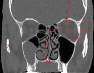


Paranasal Sinuses Ct Anatomy W Radiology
Nov 01, 12 · The entire subject of anatomy of paranasal sinuses has been rewritten after endoscopes were started to be used commonly Development of maxillary sinus CT scan axial cut showing onodi cell"Normal CT Anatomy of the Paranasal Sinuses and Nasal cavity" lecture Case discussion (901 min webinar) WEEK 2 // CT Anatomy and normal variants of PNS Reporting on selected cases (67 hour learning time) 9am CET // Live webinar "Anatomy of the Ostiomeatal Complex" lecture Case discussion (901Key words paranasal sinus;


Ct Scan Of Paranasal Sinuses Anatomy Youtube


Ct Sinus Anatomy Anatomy Drawing Diagram
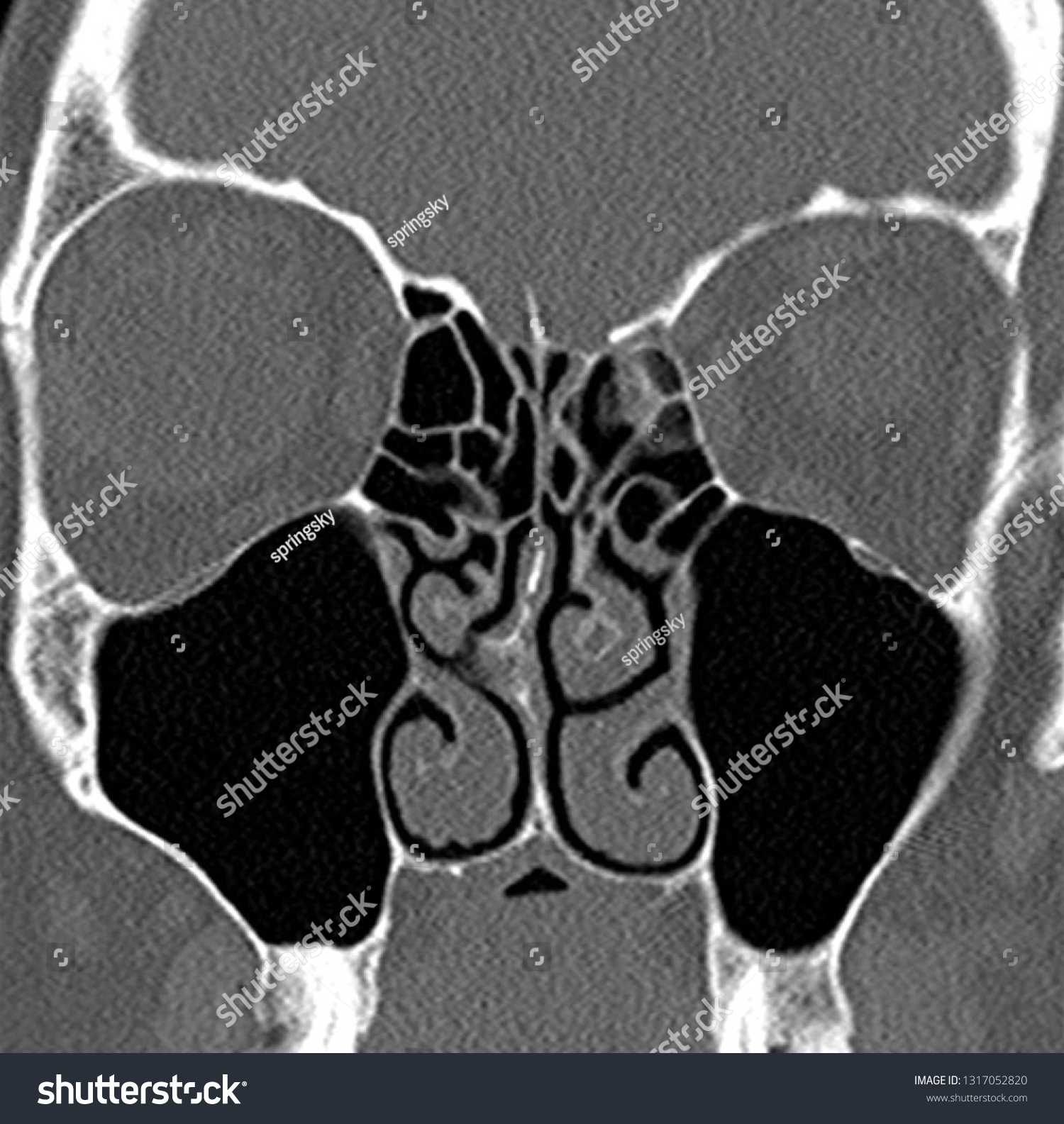
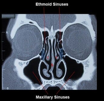
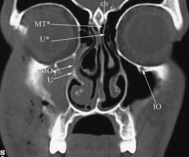
Coronal CT is usually the most functional projection for evaluating the paranasal sinuses The osteomeatal complex represents a common drainage point for the anterior sinuses (frontal, ethnoid, and maxillary) A lesion placed at the osteomeatal complex can strategically obstruct the anterior sinusesWelcome to Interactive CT Sinus Anatomy Imaging the paranasal sinuses is routine in clinical practice to evaluate for various sinus pathology, nonspecific facial pain, and preoperative planning for functional endoscopic sinus surgery (FESS), including postoperative followupSep 08, 11 · Computer tomography (CT) scanning of the paranasal sinus is one of the major modalities of investigating paranasal sinus disease Therefore, familiarisation with the radiological anatomy of this region is vital in order to perform FESS procedures safely and to preempt complications This article addresses this issue from a novice's perspective
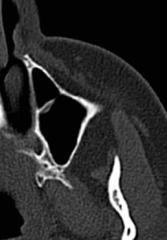


Paranasal Sinus Anatomy What The Surgeon Needs To Know Intechopen


Ct Brain And Paranasal Sinuses Impression Chronic Sinusitis Of Lt Maxillary Sinus Stock Photo Download Image Now Istock
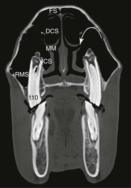
The palatine sinus is located in the hard palate There are three conchal sinuses located inside three conchae of nasal cavity The dorsal, ventral ,middle conchal sinuses located inside dorsalI want to work through the anatomy of the head as seen on CT imaging sections, but there's a lot to look at Let's start by seeing if we can recognise the frCT of Anatomic Variants of the Paranasal Sinuses and Nasal Cavity Poor Correlation With Radiologically Significant Rhinosinusitis but Importance in Surgical Planning AJR Am J Roentgenol 15 Jun;4(6) doi /AJR
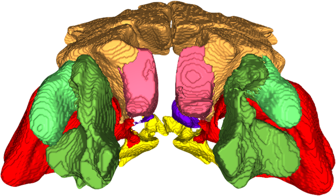


Volumetric Measurements Of Paranasal Sinuses And Examination Of Sinonasal Communication In Healthy Shetland Ponies Anatomical And Morphometric Characteristics Using Computed Tomography Bmc Veterinary Research Full Text


Startradiology
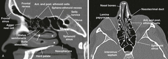
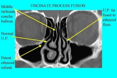
Pathology Of primary concern to the radiologist evaluating the paranasal sinuses and nasal fossae is identification of any osseous changes or variations, noting the presence of abnormal softtissue disease and its possible extension beyond the sinonasal cavities, and the characterization of thisComputed tomography (CT) of paranasal sinuses is extremely helpful for the examination of this inaccessible area Coronal CT sections of paranasal sinuses are particularly useful for surgical anatomy, as these images show nearly the same regions as the endoscopic examinationsComputed tomography (CT) scanning of the paranasal sinuses provides valuable information in assessing extent of disease and fine detailed anatomy prior to endoscopic sinus surgery Awareness of the different anatomic variants of the bony sinonasal anatomy will help the rhinologic surgeon's orientation during the procedure
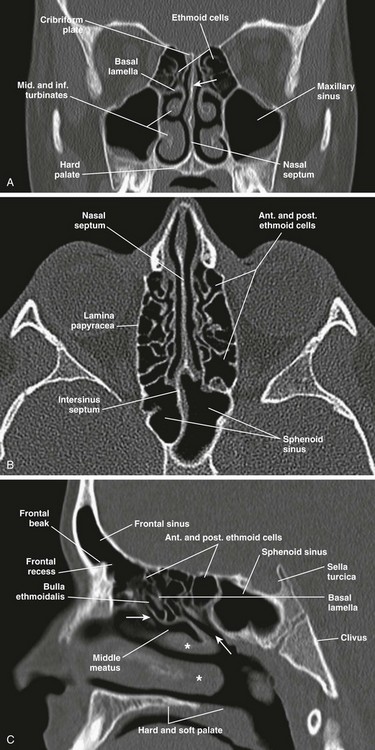


Nose And Sinonasal Cavities Radiology Key


Coronal Paranasal Sinuses
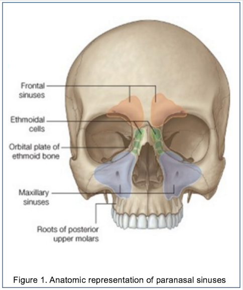
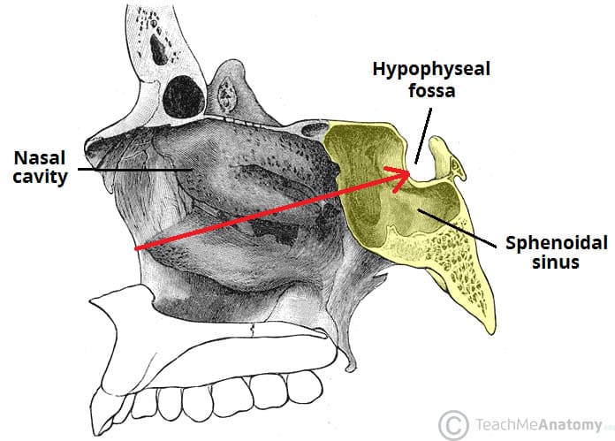
The paranasal sinuses usually consist of four paired airfilled spaces They have several functions of which reducing the weight of the head is the most important Other functions are air humidification and aiding in voice resonance They are named for the facial bones in which they are located maxillary sinus;Jul 01, 19 · Sphenoid sinuses CT brain (bone windows) The sphenoid sinus can be single or septated into multiple separate sinuses;Sep 19, 16 · The paranasal sinuses consist of paired frontal, ethmoid, maxillary, and sphenoid sinuses The frontal sinuses are located superior to the orbits and ethmoid sinuses Both the superior and posterior walls of the frontal sinus separate the sinus from the cranial vault The floor of the frontal sinus consists of the orbital roof
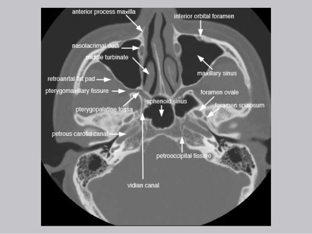


Cross Sectional Anatomy Of Paranasal Sinus


Imaging Of The Paranasal Sinuses And Nasal Cavity Normal Anatomy And Clinically Relevant Anatomical Variants Sciencedirect
The ct test is usually made to evaluate the anatomy of the paranasal sinuses Imaging the paranasal sinuses is routine in clinical practice to evaluate for various sinus pathology non specific facial pain and pre operative planning for functional endoscopic sinus surgery fess including post operative follow upComputed tomography (CT) scans may help examine paranasal sinuses' anatomy and detect abnormalities(10) Moreover, a CT scan gives a greater definition of the sinuses(11) It is more sensitive than typical radiography tools in detecting sinus pathology, especially within the sphenoid and ethmoid sinusesJan 22, 18 · Paranasal sinuses are air filled spaces located within the bones of the skull and face The sinuses are named for the bones in which they are located WATERS VIEW CALDWELLS VIEW LATERAL VIEW CALDWELLS VIEW CT IMAGES OF PARANASAL SINUSESIN DIFFERENT SECTIONS ANATOMIC VARIANTS OF PARANASAL SINUSES THANK YOU


1


Plos Neglected Tropical Diseases Facial Structure Alterations And Abnormalities Of The Paranasal Sinuses On Multidetector Computed Tomography Scans Of Patients With Treated Mucosal Leishmaniasis
The paranasal sinuses are joined to the nasal cavity via small orifices called ostiaThese become blocked easily by allergic inflammation, or by swelling in the nasal lining that occurs with a coldIf this happens, normal drainage of mucus within the sinuses is disrupted, and sinusitis may occur Because the maxillary posterior teeth are close to the maxillary sinus, this can also causeAug 08, 16 · A CT Imaging Study of the Spatial Arrangement of the Paranasal Sinuses An important prerequisite for safe endoscopic surgery within the paranasal sinuses is a clear threedimensional mental picture of their spatial relationships to each other and to the neighboring maxilla, orbit, sphenoid sinus, and anterior cranial fossaAug , 19 · The paranasal sinuses are broadly divided into two major groups the anterior sinuses and the posterior sinuses The anterior sinuses comprise the frontal sinus, anterior ethmoid air cells, and maxillary sinus These drain into a common area centered in the middle meatus, the osteomeatal unit (OMU)


Ct Scan Of Paranasal Sinus Axial Coronal Youtube


Imaging The Paranasal Sinuses Where We Are And Where We Are Going Fatterpekar 08 The Anatomical Record Wiley Online Library
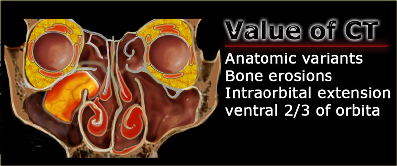
MRI examination Radiology department of the University of Pennsylvania, USA and the radiology department the Medical Centre Alkmaar, the Netherlands This article is based on a presentation given by Laurie Loevner and adapted for the Radiology Assistant by Jennifer Bradshaw This review focusses on the complimentary roles that CTAnalysis of every routine CT scan of the paranasal sinuses obtained for sinusitis or rhinitis for the presence of different anatomic variants is of questionable value unless surgery is planned Keywords anatomic variants, CT, functional endoscopic sinus surgery, paranasal sinuses, sinusitisJun 14, 17 · Performing a smooth and clean sinus surgery goes hand in hand with a perfect understanding of the nasal and paranasal anatomy Within this chapter, the paranasal and related structures surgical anatomy will be extensively reviewed, with emphasis on the anatomical landmarks and the normal anatomical variations, which have a significant impact on the function,
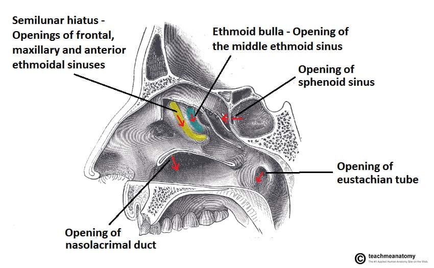


The Paranasal Sinuses Structure Function Teachmeanatomy
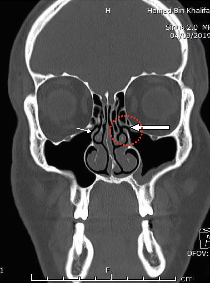


Radiology Of Paranasal Sinuses Springerlink
The ct test is usually made to evaluate the anatomy of the paranasal sinuses The module interface is meant to mimic a radiology workstation with adjacent image scrolling via arrow keys and or mouse wheel button Straps and pillows may be used to help the patient maintain the correct position and to hold still during the examHuman Senses by Dr Gregorius Enrico, SpRad Anatomy by Dr Namit Mathur SENOS PARANSALES by Oswaldo Tostado Islas OMFS EXAM by Dr ANKUSH Skull and facial bone anatomy by Andrew Murphy Sinuses by Luca Paul Milford CT anatomi by Tunjay Mammadov Anatomy H&N by Dr Phillip Marsh Annotated anatomy by Dr Sachintha HapugodaFeb 09, 21 · Anatomy of the head on a cranial CT Scan brain, bones of cranium, sinuses of the face Coronal Brain CT Vasculary territories Dural venous sinuses, Veins, Arteries Bones of cranium Axial CT Paranasal sinuses CT Cranial base , CT Foramina, Nasal cavity, Paranasal sinuses Bones of cranium Anatomy , CT Invalid input


Anatomy Ento Key
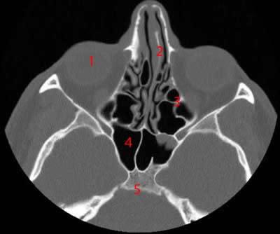


Paranasal Sinuses Ct Anatomy W Radiology
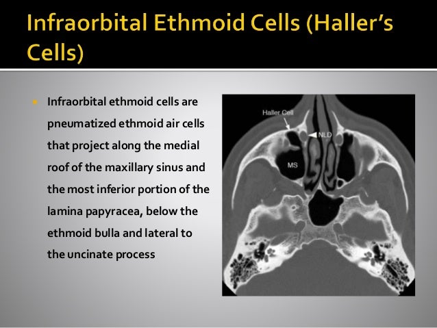
Numerous sinonasal anatomic variants exist and are frequently seen on sinus CT scans The most common ones are Agger nasi cells, infraorbital ethmoidal (Haller) cells, sphenoethmoidal (Onodi) cells, nasal septal deviation, and concha bullosa 1 – 10 The Agger nasi cells are the most anterior ethmoidal air cellsMar 01, 17 · The paranasal sinuses are group of air filled spaces surrounding the nasal cavity Paranasal sinuses start developing from the primitive choana at 25–28 weeks of gestation Three projections arise from the lateral wall of the nose and serve as the beginning of the development of the paranasal sinusesOct 13, 14 · 63 CT is currently the modality of choice in the evaluation of the paranasal sinuses and adjacent structures Its ability to optimally display bone, soft tissue, and air provides an accurate depiction of both the anatomy and the extent of disease in and around the paranasal sinuses In contrast to standard radiographs, CT clearly shows the fine bony anatomy of the


Prognostic Value Of Sinus Ct Scans In Hematopoietic Stem Cell Transplantation Brazilian Journal Of Otorhinolaryngology


Pdf Ct Of The Paranasal Sinuses Normal Anatomy Variants And Pathology
May 18, 21 · CT anatomy of the nasal cavity and paranasal sinuses is described in detail together with the anatomic variants encountered in each region Imaging techniques and protocol Preoperative CT imaging of the paranasal sinuses is performed after completion of the medical treatment because up to 80% of patients suffering from acute upper respiratory tract infectionComputed tomographic (CT) scanning of the face has become a standard part of oromaxillofacial imaging Variations in paranasal sinus anatomy as shown on CT scans is of potential significance, for it may pose risks during surgery or predispose to certain pathologic conditions and diseases Studying the relative frequency and concurrence ofFeb 25, 09 · Role of CT and MR When it comes to imaging of neoplasms of the paranasal sinuses, CT and MRI play complementary roles It is not about the histology but about answering the question 'is it tumor or not?' and then determining the extent of the disease, for example intracranial or orbital extension


Startradiology


A E Coronal Axial And Sagittal Ct Images Of The Bone Window Download Scientific Diagram


Variability Of Paranasal Sinus Pneumatization In The Absence Of Sinus Disease Ochsner Journal


Ct Anatomy Of The Paranasal Sinuses


Pin On Anatomia


Maxillary Sinus Radiology Reference Article Radiopaedia Org


Role Of Anatomic Variations Of Paranasal Sinuses On The Prevalence Of Sinusitis Computed Tomography Findings Of 350 Patients Kaya M Cankal F Gumusok M Apaydin N Tekdemir I Niger J Clin Pract


Figure 5 From Paranasal Sinus Anatomy What The Surgeon Needs To Know Semantic Scholar


The Radiology Assistant Mri Examination


Nose And Sinonasal Cavities Radiology Key


Nasal Cavity Anatomy Physiology And Anomalies On Ct Scan Overview Anatomy And Physiology Of The Nasal Cavity Nasal Cavity Anomalies And Sinusitis


Paranasal Sinus Anatomy What The Surgeon Needs To Know Intechopen


Computed Tomography Anatomy Of The Paranasal Sinuses And Anatomical Variants Of Clinical Relevants In Nigerian Adults Sciencedirect


Http Pdf Posterng Netkey At Download Index Php Module Get Pdf By Id Poster Id


The Radiology Assistant Paranasal Sinuses Mri


Epos C 2117


Ct And Mri Show Complexparanasal Sinus Anatomy


Basic Ct Anatomy Of Paranasal Sinuses Made Easy Youtube


Pictorial Essay Anatomical Variations Of Paranasal Sinuses On Multidetector Computed Tomography How Does It Help Fess Surgeons Abstract Europe Pmc


Ct Of Paranasal Sinuses Stock Image P410 0085 Science Photo Library


Pin On Anatomia


The Paranasal Sinuses Structure Function Teachmeanatomy


The Preoperative Sinus Ct Avoiding A Close Call With Surgical Complications Radiology


1


The Preoperative Sinus Ct Avoiding A Close Call With Surgical Complications Radiology


Www Ajronline Org Doi Pdf 10 2214 Ajr 14


Pns Ct Scan Anatomy Ct Scan Machine


Crista Galli Pneumatization Is An Extension Of The Adjacent Frontal Sinuses American Journal Of Neuroradiology


Ct Signs Of Pressure Induced Expansion Of Paranasal Sinus Structures


Ct Scan Human Paranasal Sinuses Many Stock Photo Edit Now


Unusually Large Radicular Cyst Presenting In The Maxillary Sinus Bmj Case Reports


Cureus Paranasal Sinus Wall Erosion And Expansion In Allergic Fungal Rhinosinusitis An Image Scoring System



Ct And Mr Anatomy Of Paranasal Sinuses Key Elements Springerlink


Sinus Anatomy Sinushealth Com


Paranasal Sinuses Annotated Ct Radiology Case Radiopaedia Org
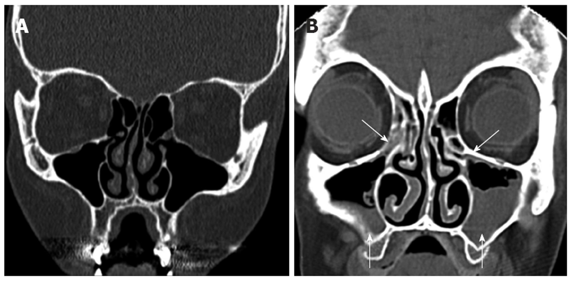


Computed Tomography Scans Of Paranasal Sinuses Before Functional Endoscopic Sinus Surgery


Startradiology


Nasenhhle Nasennebenhhlen Ct Nasal Cavity Paranasal Sinuses Tillman


The Frontal Sinus Drainage Pathway And Related Structures American Journal Of Neuroradiology


Www Ijcmr Com Uploads 7 7 4 6 Ijcmr 1967 V4 Pdf


Pin On Paranasal Sinuses


Multiplanar Computed Tomographic Analysis Of Frontal Cells According T Rmi


Ct Signs Of Pressure Induced Expansion Of Paranasal Sinus Structures


Paranasal Sinus Anatomy What The Surgeon Needs To Know Intechopen
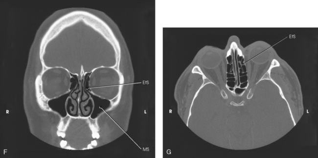


Paranasal Sinuses Radiology Key
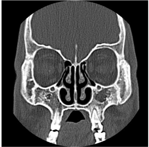


Paranasal Sinus Ct Coronal Section Indicating Aplasia Open I


Ejo Springeropen Com Track Pdf 10 4103 1012 5574


Epos C 093


Http Pdf Posterng Netkey At Download Index Php Module Get Pdf By Id Poster Id


Home Page


Normal Anatomy And Anatomic Variants Of The Paranasal Sinuses On Computed Tomography Neuroimaging Clinics


Computed Tomography Anatomy Of The Paranasal Sinuses And Anatomical Variants Of Clinical Relevants In Nigerian Adults Sciencedirect


1
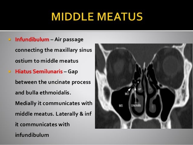


Ct Anatomy Of Para Nasal Sinuses


Reducing The Radiation Dose For Low Dose Ct Of The Paranasal Sinuses Using Iterative Reconstruction Feasibility And Image Quality European Journal Of Radiology


Computed Tomography Anatomy Of The Paranasal Sinuses And Anatomical Variants Of Clinical Relevants In Nigerian Adults Sciencedirect


Anatomic Variations Of The Paranasal Sinuses Ct Examination For Endoscopic Sinus Surgery Auris Nasus Larynx


Paranasal Sinus Disease Ento Key


The Preoperative Sinus Ct Avoiding A Close Call With Surgical Complications Radiology


Startradiology


The Canine Head And Skull Ct Atlas Of Veterinary Clinical And Radiological Anatomy Of The Dog
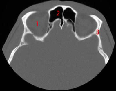


Paranasal Sinuses Ct Anatomy W Radiology
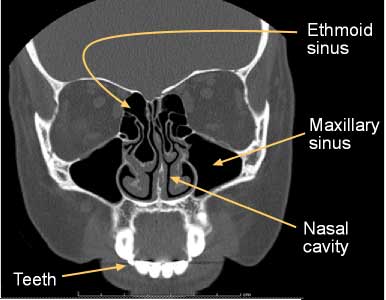


Computed Tomography Ct Sinuses


A Ct Scan In Coronal Plane Shows The Normal Paranasal Sinus Anatomy Download Scientific Diagram


Ct Scan Anatomy Of Paranasal Sinuses Professor Dr Muhammad Ajmal Ppt Video Online Download


Ct Scan Anatomy Of Paranasal Sinuses Professor Dr Muhammad Ajmal Ppt Video Online Download


Ct Sinuses Anatomy Quiz Radiology Case Radiopaedia Org


Axial Ct Scan Of The Nose And Paranasal Sinuses Showing That The Lesion Download Scientific Diagram


Coronal Paranasal Sinuses
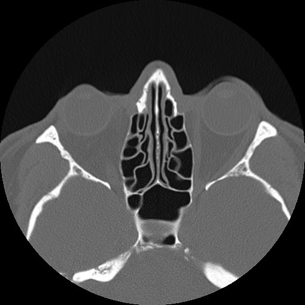


Normal Sinus Ct Annotated Radiology Case Radiopaedia Org


Anatomical Variations Of Nose And Para Nasal Sinuses Ct Scan Review In South Gujarat Semantic Scholar


Normal Ct Paranasal Sinuses Radiology Case Radiopaedia Org
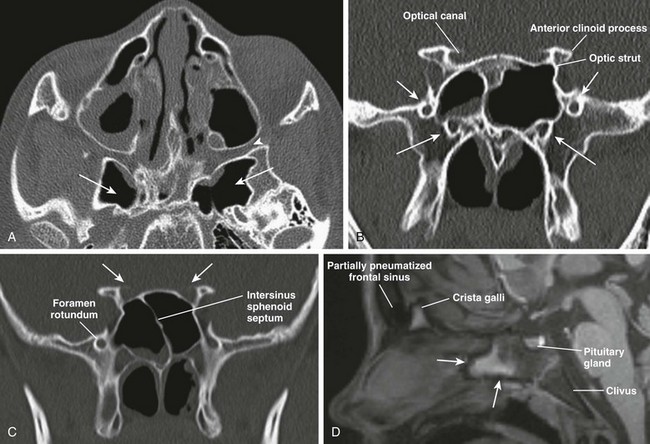


Nose And Sinonasal Cavities Radiology Key


Agenesis Of Paranasal Sinuses And Nasal Nitric Oxide In Primary Ciliary Dyskinesia European Respiratory Society


Ct Scan Of The Paranasal Sinuses History Basic Concepts Anatomy


Q Tbn And9gctc4evwb3vmd0f Phr3hylwnpjlosti9 Rvyip5jrwyk4w8wf0l Usqp Cau
:background_color(FFFFFF):format(jpeg)/images/article/en/the-paranasal-sinuses/972PC0nYOzlz7wqSgLmNA_sinus_frontalis_large_u9Vfozc0uUoMtc6KtIaUfw.png)


Paranasal Sinuses Anatomy And Clinical Aspects Kenhub


Imaging The Paranasal Sinuses Where We Are And Where We Are Going Fatterpekar 08 The Anatomical Record Wiley Online Library


Imaging Of Paranasal Sinuses And Anterior Skull Base And Relevant Anatomic Variations Radiologic Clinics


Diseases Of The Nasal Cavity And Paranasal Sinuses Veterian Key


Imaging The Paranasal Sinuses Where We Are And Where We Are Going Fatterpekar 08 The Anatomical Record Wiley Online Library


The Preoperative Sinus Ct Avoiding A Close Call With Surgical Complications Radiology


コメント
コメントを投稿